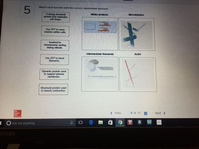
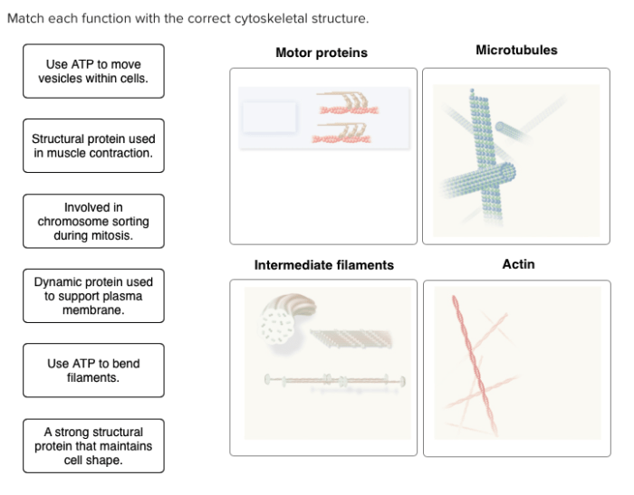
Match each function with the correct cytoskeletal structure. The cytoskeleton, a complex network of protein filaments and tubules, plays a pivotal role in maintaining cell shape, facilitating cell division, transporting organelles, and enabling muscle contraction. Understanding the relationship between cytoskeletal structures and their functions is crucial for unraveling the intricate workings of cells.
This comprehensive guide delves into the three main cytoskeletal structures: microtubules, microfilaments, and intermediate filaments. We will explore their distinct compositions, diameters, and the specific functions they perform within the cell.
Cytoskeletal Structures: Match Each Function With The Correct Cytoskeletal Structure.

The cytoskeleton is a complex network of protein filaments that extends throughout the cytoplasm of eukaryotic cells. It provides structural support, maintains cell shape, and facilitates various cellular processes such as cell division, organelle transport, and muscle contraction.
Microtubules, Match each function with the correct cytoskeletal structure.
Microtubules are long, hollow cylinders composed of tubulin protein subunits. They are the thickest cytoskeletal structures, with a diameter of approximately 25 nanometers. Microtubules play a crucial role in cell division, forming the mitotic spindle that separates chromosomes during mitosis and meiosis.
They also participate in organelle transport, serving as tracks along which motor proteins move organelles throughout the cell.

Microfilaments
Microfilaments are thin, solid filaments composed of actin protein subunits. They have a diameter of approximately 7 nanometers and are responsible for maintaining cell shape and facilitating muscle contraction. Microfilaments form a meshwork beneath the plasma membrane, providing structural support and allowing cells to change shape during processes like cell division and movement.

Intermediate Filaments
Intermediate filaments are intermediate in size between microtubules and microfilaments, with a diameter of approximately 10 nanometers. They are composed of a diverse group of proteins and provide mechanical support to cells. Intermediate filaments form a network throughout the cytoplasm, helping to maintain cell shape and resist mechanical stress.

Matching Functions to Structures
| Function | Structure 1 | Structure 2 | Structure 3 |
|---|---|---|---|
| Cell shape maintenance | Microfilaments | Intermediate filaments | |
| Cell division | Microtubules | ||
| Organelle transport | Microtubules | ||
| Muscle contraction | Microfilaments |
Key Questions Answered
What is the primary function of microtubules?
Microtubules are responsible for maintaining cell shape, facilitating cell division, and transporting organelles within the cell.
How do microfilaments contribute to cell movement?
Microfilaments play a crucial role in cell shape maintenance and muscle contraction.
What is the unique characteristic of intermediate filaments?
Intermediate filaments provide mechanical support to cells and contribute to cell shape maintenance.